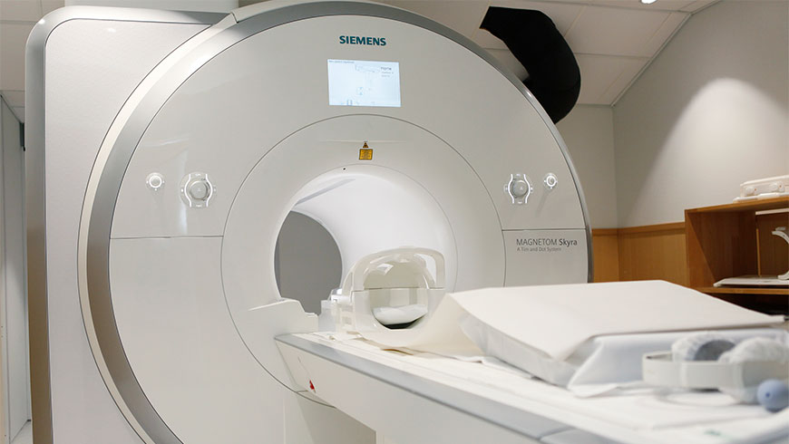
The Mehiläinen Imaging Units offer a wide range of radiological examinations, including
- Various native radiogram examinations◦with referral from your doctor, no appointment is needed
- Mammograms, ultrasound examinations◦with referral from your doctor, by appointment
- Computer tomography (CT)◦with referral from your doctor, by appointment (available only at Mehiläinen Hospital Töölö)
- Cone-beam computer tomography (CBCT) examinations of the limbs and parasanal sinuses◦with referral from your doctor, by appointment
- Biopsies and injections with ultrasound control◦with referral from your doctor, by appointment
- Echocardiograms◦by appointment with our cardiologist consultants
- MRI examinations◦with referral from your doctor, by appointment (only available at Mehiläinen Hospital Töölö)
Magnetic resonance imaging
Mehiläinen offers magnetic resonance examinations to private and occupational healthcare customers of all ages. In magnetic resonance imaging, an intense magnetic field together with radio wave impulses help us obtain clear anatomic images of the area to be examined. The examination is harmless and pain-free to the patient, and it is carried out without using radiation. The procedure takes c. 45 minutes, and the patient must lie still during it so that a good image quality is ensured.
During a magnetic resonance examination, the patient lies still on his/her back inside an imaging machine. During imaging, there will be loud noises from the machine. Protective ear phones will be provided with an option to listen to music. Precise images of the area under examination will be taken in two or three perpendicular planes. For some examinations, examination reels are used that receive target-specific image signals and these are positioned above the area to be examined. Using magnetic resonance imaging, diseased and healthy tissue can be easily distinguished.
Magnetic resonance imaging can be used to examine the areas of the head, joints and limbs, neck, chest, lumbosacral (spine), lungs, breasts, abdomen and pelvis. In addition, blood vessels can be examined either with or without the use of a contrast agent. In some examinations contrast agent is injected intravenously or inside a joint in order to identify potential changes in the area under examination.
Mammography
With a doctor's referral, Mehiläinen offers mammogram and breast ultrasound examinations. In general, so called extensive X-ray mammography together with an ultrasound scan of the breasts are carried out at Mehiläinen. Mammography is a traditional imaging method using X-ray radiation, which is used for investigating the diseases of the mammary glands. In most cases, an ultrasound examination of the breasts is added to a mammogram as these two investigative methods complement each other. Some changes are most reliably detected using a mammogram, whilst others are most obvious in an ultrasound scan.
During an X-ray examination, the radiographer in general takes mammogram images from two different directions. If ifrequired, targeted or magnified images can be taken in order to evaluate minor changes. During the imaging, inside the imaging device, the breast to be examined will be pressed evenly between a plastic plate and the imaging cassette so as to improve picture quality and to minimise the radiation stress caused by the imaging.
A mammogram and a breast ultrasound scan usually takes a maximum of 30 minutes. Based on preliminary information provided by the referring doctor in his/her examination request, the imaging results and any possible comparisons that can be made with previous images, the radiologist will provide a written report on the examination.
X-ray
With a doctor's referral, Mehiläinen offers X-ray examinations to private and occupational healthcare customers of all ages.
An X-ray examination is a traditional imaging method using X-rays, which is used in the examination of a variety of diseases and traumas to the locomotor system, respiratory system and cardiovascular system. Nowadays, X-ray examinations are less often used for examinations of the abdominal area and the urinary tract.
During an X-ray examination, a radiographer takes the traditional X-ray images of the area to be examined from a minimum of two different directions. For the spine and some joints, images will often even be taken from several directions. The imaging will be carefully limited to the area to be examined. It is important not to move during the imaging. The procedure generally takes some minutes.
Common reasons for having an X-ray are respiratory inflammations, such as suspected sinusitis or pneumonia, the evaluation of the degree of cardiac insufficiency as well as any degenerative, inflammatory or accident damage to bones and joints.
Based on preliminary information provided by the referring doctor in his/her examination request, the imaging results and any possible comparisons that can be made with previous images, the radiologist will provide a written report on the examination.
Computerised Tomography
Computerised tomography (CT) is an X-ray examination, which provides cross-sectional images of the body. This examination is quick and easy and during it the patient lies on the examination table, which passes through a ring-like imaging machine. A CT scan is well suited to those patients who suffer from claustrophobia.
Ultrasound
With a doctor's referral, Mehiläinen offers ultrasound examinations to private and occupational healthcare patients of all ages.
An ultrasound examination is an established imaging method, which uses sound waves within the ultrasound range to form an image in a similar way to an echo sounder. Ultrasound is used for the examination of a variety of diseases of and traumas to the locomotor system as well as for examination of the diseases of the cardiovascular system. Ultrasound is often used as a standard test during any examination of the abdominal organs as well as the urinary tract, and it can also be used to study many subsurface soft tissue objects.
At the imaging unit, ultrasound examinations are carried out by a radiologist. A cardiologist generally performs ultrasound scans of the heart; urologists and gynaecologists also carry out ultrasound examinations on patients in their own area of speciality.
Customary reasons for an ultrasound examination are e.g. suspected gall stones, various kidney diseases, accidents to and diseases of the ligaments, tendons and muscles, as well as lumps in the breasts and the neck area. During the examination, needle biopsies may be taken as and when required.
Based on preliminary information provided by the referring doctor in his/her examination request, the imaging results and any possible comparisons that can be made with previous images, the radiologist will provide a written report on the examination.